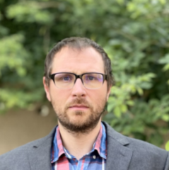
Douglas Shepherd
Seminar Information

Continued advancements in optical microscopy and molecular labeling techniques have enabled the multi-dimensional visualization of biology in action, with measurements now spanning from individual molecules through organism behavior. However, the quality and confidence of biological and biophysical knowledge extracted from imaging experiments depend on the molecular labeling strategy, instrument design, and detector choices. At the molecular and macromolecular level, compromises are often necessary to achieve the required optical resolution, volume size, imaging speed, number of molecules, samples, or other biologically driven experimental design criteria. Unfortunately, such compromises inevitably increase uncertainty when quantifying molecular identity, dynamics, and interactions. In this talk, I will discuss our recent efforts to develop new fast and scalable 3D imaging approaches that break through the existing space-time bandwidth barriers. I will focus on label-free, quantitative phase measurements using single-shot optical diffraction tomography at >1,000 volumes per second and fluorescence imaging across time and length scales using high-resolution oblique plane microscopy.
Douglas Shepherd is an assistant professor in the Center for Biological Physics and the Department of Physics and a member of the Center for Mechanisms of Evolution and Neuroscience Program at Arizona State University. He is currently a Scialog Advanced Bioimaging fellow and directs the Quantitative Imaging and Inference Laboratory (qi2lab). The qi2lab develops and applies quantitative imaging methods to understand self-organization and cellular “decision making” in the context of the physical principles that govern the microscopic world.
